Researchers from the University of Westminster have demonstrated that subtle changes in muscle shape, detectable through advanced MRI analysis, can provide important insights into metabolic health, ageing and the risk of type 2 diabetes. The research has gained national media coverage and was presented at the Radiological Society of North America’s (RSNA) Annual Meeting in Chicago.
Led by Dr Marjola Thanaj and Professor Louise Thomas from the Research Centre for Optimal Health (ReCOH), the study focused on the gluteus maximus, one of the largest and most metabolically important muscles in the human body. By analysing tens of thousands of MRI scans using three-dimensional MRI shape mapping, the researchers showed that alterations in muscle shape rather than simple loss of volume are associated with ageing, lifestyle factors, osteoporosis and type 2 diabetes, with clear differences observed between men and women.
The work draws on large-scale imaging data from UK Biobank’s record-breaking human imaging project, which Professor Thomas has worked on closely for the past 15 years, and showcases how next-generation image analytics can uncover clinically meaningful biomarkers beyond traditional measures such as muscle size or body weight.
Dr Thanaj and Professor Thomas worked with Westminster colleagues Dr Brandon Whitcher, Camilo Bell-Bradford, Dr Marili Niglas and Professor Jimmy Bell in collaboration with muscular skeletal radiologist Dr Dimitri Amaras and Hamzah Raza from Imperial College London.
About the research project Dr Thanaj said: “Unlike past studies that mainly looked at muscle size or fat, we used 3D shape mapping to pinpoint exactly where the muscle changes, giving a much more detailed picture. People with higher fitness, as measured by vigorous physical activity and hand grip strength, had a greater gluteus maximus shape, while aging, frailty and long sitting times were linked to muscle thinning. These findings suggest that muscle shape may serve as a sensitive imaging-derived biomarker of metabolic and functional health, potentially enabling earlier risk stratification than current approaches.”

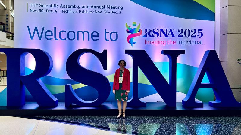
Professor Thomas at the RSNA conference
The research was recently presented at the Radiological Society of North America’s (RSNA) Annual Meeting in Chicago and attracted significant interest due to its implications for personalised medicine and preventive healthcare. The study has also received widespread media attention, including national press coverage and an interview with Professor Thomas on BBC Radio 4 Woman’s Hour, reflecting its relevance to both clinical practice and public health.
Professor Thomas was also invited by the RSNA to speak at a session sponsored by their Public Information Committee titled Obesity: More than Meets the Eye - the Role of the Radiologist. Her talk focused on multiorgan imaging in obesity which also used data from the UK Biobank.

Professor Thomas delivering a talk on multiorgan imaging in obesity
About the conference Professor Thomas said: “RSNA is the biggest radiology conference in the world with over 50,000 people attending. The technical exhibition is fantastic and a great insight into the direction of the field from an industrial perspective. It was a huge honour for me to be invited back to speak again this year and very exciting that our work on the gluteus maximus was so prominently featured.”
This research project directly contributes to the United Nations Sustainable Development Goals (SDGs) 3: Good Health and Wellbeing and 17: Partnerships for the Goals. Since 2019, the University of Westminster has used the SDGs holistically to frame strategic decisions to help students and colleagues fulfil their potential and contribute to a more sustainable, equitable and healthier society.
Learn more about Biological and Biomedical Sciences courses at the University of Westminster.
Find out more about Westminster’s Research Centre for Optimal Health.
Related articles


