Dr Nicolas Basty and colleagues from the University of Westminster’s Research Centre for Optimal Health have published a new paper in the journal Scientific Reports, in collaboration with scientists from Calico Life Sciences about the use of MRI data to measure core muscles.
The paper explains how they have used MRI data from the UK Biobank, a major national and international health resource and registered charity, in combination with a newly developed machine learning model for the automated measurement of the iliopsoas muscles, or ‘core-muscles’ as they are more commonly known, which are a key indicator of frailty. The study found that muscle decline was more prominent in older individuals, especially in males over 60 years of age.
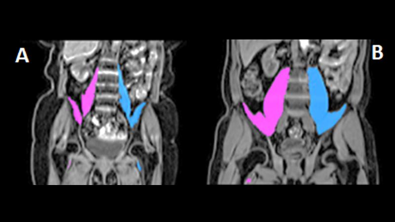
The iliopsoas muscles are found in the abdomen and pelvis and are extremely important as they are used in almost every day-to-day activity, from posture to walking and running. They are involved in moving the hips and keeping the spine stable. Their size can indicate the state of someone’s health – small iliopsoas muscles are associated with frailty and can predict outcomes and survival from surgery and disease, whereas larger iliopsoas muscles are linked with health and fitness.
Until now, most studies have measured iliopsoas size manually and in 2D, which is a time-consuming and error prone method. In this study, the team developed a machine learning model to automatically measure the 3D volume of the iliopsoas muscles from MR images in seconds. The model took less than a day to predict the results for 5,000 people.
Talking about the research, Dr Basty said: “Thanks to machine learning and the availability of large amounts of medical imaging data from the UK Biobank, we are able to predict those muscle measurements in thousands of people in order to investigate the effects of health, ageing, and disease on those essential muscles.
“We look forward to performing further analysis of this particular measurement, as well as our many other currently ongoing projects. We are very fortunate to be in a position where we are able to carry out our work fully remotely, thanks to daily video meetings, remote access to our powerful computers, not to forget the hard work from the people at the UK Biobank in data acquisition, and most of all the wonderful volunteers.”
Professor Louise Thomas, Professor of Metabolic Imaging at the University of Westminster, added: “This work has shown that not only is it possible to automatically measure the size of the iliopsoas muscle in a very large cohort, but that these measurements can be made very accurately. Undertaking this sort of analysis manually would not be feasible in such a large study.
“This advance has enabled us for the first time to establish normal population reference values and understand how the iliopsoas varies in size and is influenced by factors such as height, weight, age and gender. Going forward, we intend to expand this work to include a much larger cohort, around 40,000 people, and investigate genetic factors which may play a role in determining iliopsoas size and related health outcomes.”
Read the full study in the journal Scientific Reports.
Find out more about the Research Centre of Optimal Health at the University of Westminster.
Related articles


